More Imaging Modalities
Color Maps
How do we relate a pixel to a certain brightness or color?
Anatomical Vs. Molecular Image
Anatomical shows cell parts and body parts, molecular shows the concentration or fluorescent markers, etc.
Molecular Image
An image where each pixel presents gene expression or protein concentration.
-Paint molecular information on top of anatomical image
-Color is proportional to molecular concentration
-Paint molecular information on top of anatomical image
-Color is proportional to molecular concentration
Image Generation
detect beacons in the body by the detector on the imaging modality.
x-Rays
Form of electromagnetic radiation with wavelengths SHORTER than visible light (shorter wavelength = higher frequency)
CT Scans
Computed Tomography
-3D form of X-Rays
-The X-Ray beam has to move all around the patient and scan from hundreds of different angles
Detects STRUCTURE
-3D form of X-Rays
-The X-Ray beam has to move all around the patient and scan from hundreds of different angles
Detects STRUCTURE
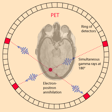
PET Scan
Positiron Emission Tomography
-Require a contrast agent
-In a PET scan, patient s injected with radioactive substance and placed on a flat table that moves in increments through a donut shaped housing
-housing contains the circular gamma ray detector array, which has a series of scintillation crystals, each connected to a photomultiplier tube.
Detects FUNCTION
Ultrasounds
uses high frequency sound waves and their echoes
Example
Use MRI as the primary imaging modality
-molecular nanosystems will manipulate the magnetic nanoenvironment
-molecular nanosystems will manipulate the magnetic nanoenvironment
Image
Can be 2D or 3D, where the arguements of the 2D function are integers x(n,m)
Detection
Detector detects brightness or color, and each pixel is given a certain "value". The pixels that have the value will be a certain color and the pixels that don't will not.
Detection
HIgher concentration = higher value for the pixel = higher brightness/harder color
Overlay
Take the color map and overlay it on the anatomical image to see the concentration of the particular molecule in that pixel.
Creating Color Maps
The color/brightness assigned to a pixel depends on the value of the colormap function
- the simplest function is X(n,m) == C*(X/a)
where
C = index value, based on color palate
a = scale factor, based on maximum [target molecule]
x = [target molecule]
- the simplest function is X(n,m) == C*(X/a)
where
C = index value, based on color palate
a = scale factor, based on maximum [target molecule]
x = [target molecule]
Other Imaging Technologies
Description
Note
Description
Optical Imaging
Microscopy
Fluorescent
Fluorescent
MRI
TBDISCUSSED
Applications
Best and fastest tools for studying chest, abdomen, and pelvis because it provides detailed, cross-sectional views of all types of tissue.
- useful in detection of cancers and tumors
- diagnosis, detection, treatment of vascular disease
- Pulmonary embolisms
- Bones injuries, orthepedic injuries
- real-time guides to physicians during interventional procedures
X-Ray Generation
Generated by accelerating electrons inside a vacuum, then colliding these high velocity electrons with a metal target.
- Electrons FLY off of the cathode released from the filament, and are attracted to the anode (tungsten tube) and the electrodes hit the disk and collide with the tungston, causing x-rays to be formed.
- Electrons FLY off of the cathode released from the filament, and are attracted to the anode (tungsten tube) and the electrodes hit the disk and collide with the tungston, causing x-rays to be formed.
X-Ray Fluorescence
The high energy collisions dislodge an electron in a low-energy orbital (ionizing radiation)
-An electron from a higher energy orbital falls to the lower energy level, and releases energy in the form of a photon
-An electron from a higher energy orbital falls to the lower energy level, and releases energy in the form of a photon
Bremsstrahlung
Do not need a collision between electrons to create energy photon
- electron is attracted to the atom's nucleus
-as the electron gets close, it slows down and changes course.
-because they slow down, the electron releases energy (in the form of an x-ray photon)
- electron is attracted to the atom's nucleus
-as the electron gets close, it slows down and changes course.
-because they slow down, the electron releases energy (in the form of an x-ray photon)
Medical Imaging
The photons xrays get directed toward the patient, the body absorbs some of the x-ray photons
-the ones that pass through reach a radiographic film. If the x-rays reach the film, the film turns dark. If it does not, the film stays clear.
-the ones that pass through reach a radiographic film. If the x-rays reach the film, the film turns dark. If it does not, the film stays clear.
Avantages
You can see all the organs, sizes locations,
You can see things that are behind other things, smaller bones get hidden behind smaller bones.
You can see things that are behind other things, smaller bones get hidden behind smaller bones.
How it Works
Crystals interact with the gamma rays, and the photomultiplier tubes convert and amplify the photons to electrical signals.
- signals are processed by the computer to generate images.
- signals are processed by the computer to generate images.
Application
More targeted at detecting metabolism and functional things as oppose to CT Scans that detect structural features of the body.
PET Image
Imaging
The machine transmits high frequency sound pulses into patients body using a probe.
- Sound waves travel into the body and hit a boundary between tissues
- Some get reflected back, some travel further until another boundary is reached
- Reflected waves are picked up by the probe and relayed to the machine
- The machine calculates the distance from the probe to the tissue or organ using the speed of sound in tissue and the time of each echo's return